shaderware recommend this title as an accompaniment to virtual radiographyTM

shaderware recommend this title as an accompaniment to virtual radiographyTM
Virtual environments are now accepted as valuable for training in pilot training, driving, deep sea diving, parachute training, fire-fighting, air traffic control, power station control, ship mannovers, battlefield training, surgery, anesthetics, and space. This is because they embody many of the characteristics of the ideal medium, they are student centred and highly interactive (Psotka 1995, Schroder 1995).
Virtual training is especially valuable when the task involves danger (ionising radiation), takes place in an unpredictable environment (hospital), is difficult to timetable (large student numbers) and where alternatives are expensive (live x-ray rooms and CR/DR systems) (Rose et al. 2000) The use of virtual radiographyTM allows the lecturer or clinical mentor total control of the situation given to the student and allows comprehensive monitoring of performance. By adopting a computer game format, students have been shown increased motivation (Schroder 1995).
In the training of spatial skills positive transfer from virtual to real has been reported without exception (Regian 1992, Arthur et al. 1997, Waller et al. 1998, Brooks et al. 1999).
Reigan et al.(1992) and Brooks et al.(1999) have found clear evidence of positive transfer of procedural learning from virtual to real environments.
How does virtual radiography improve student performance? This mechanism is not certain, but a number of ways have been suggested; for example the student may gain a simple familiarity with the x-ray room and its components, which aids subsequent learning in clinical practice. Alternatively, it could be due to particular cues 'jumping out' at a student from their practices in the virtual radiography room.
The virtual radiography experience also allows students to rehearse specific sequences of actions, such as placing cassettes in the bucky and centring to them. Rose et al. (2000) also found that virtual environments help students remember things due to the mixing of 'where' and 'when'. Students learn the right thing at the right time in the right physical space, rather than reading a book or listening to a lecture or even passively watching an animation or video.
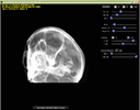
The virtual radiographyTM applications use the CT data obtained by The National Library of Medicine Visible Human Project to generate simulated radiographs based on user supplied parameters. These parameters include both geometric positioning data (e.g. patient position and orientation, tube location and angulation) and technical factors (e.g. tube voltage, current and exposure time).
The software then uses these parameters to trace the path of a virtual x-ray beam as it travels from the tube to the sensor. For a representative set of rays striking the sensor the software calculates how the original beam intensity would have been attenuated, given its individual path through the virtual human. Based on the intensity of the attenuated beam, a grayscale image is generated based on a simulated receptor response curve.
As you can imagine, the number of calculations required to build the final image is enormous, counted in the billions, is beyond the processing capabilities of the central processor used in current consumer computers. Fortunately, the impetus of computer gaming has driven the development of graphics processors to the point where these dedicated chips are extremely powerful parallel processing units. The virtual radiographyTM software uses this specialized hardware to perform the geometric, resampling and attenuation calculations in a realistic timescale. While today's top end graphics processor aimed at the gaming market will produce a simulated image in less than a second, processors sold in 2002 can produce an acceptable image in a few seconds. This means that virtual radiographyTM can be used on very modest hardware, although there is a minimum hardware requirement
At the heart of the virtual radiographyTM application is the DRR engine that generates the simulated radiographs. We have developed individual modules that are designed to model various radiographic scenarios. We currently offer the following modules
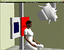
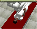
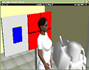
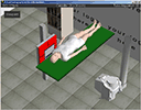
Radiographic examination of the head and neck using an erect bucky and/or cassette stand.
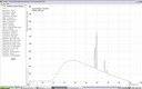
Presents a spectrum in graphical form, which changes shape instantly with any change in exposure factor or equipment choice.
Provides real time changes to the radiographic image to support lectures and workshops with larger student groups.
The recommended operating system for virtual radiographyTM is Microsoft Windows XP. The software has been tested and found to work correctly on Windows 2000 and Windows Vista. At the present time, these are the only supported operating systems.
In order to compute a simulated radiograph, billions of calculations are required. Virtual Radiography harnesses the power of the graphics processor to perform these calculations fast enough to create the image in a realistic timescale. Consequently, the most critical component of your system from the point of view of virtual radiography is the graphics processor or GPU as it is also known.
The technology that underpins virtual radiographyTM was developed to satisfy the graphics requirements of computer games. In order to run virtual radiographyTM you will not need the latest and greatest graphics processor, most chips introduced after 2003 should be adequate. For the more technically minded, the processor must be Directx v9.0c and support floating point render targets. Chips from nvidia 6000 series and better, or ATI Radeon 9600 or better will run the software. Unfortunately the majority of embeded graphics processors from Intel are not suitable.
More information can be found in the technical FAQ and the list of tested graphics cards
You can see what has been written about Virtual RadiographyTM in the press but we feel the only way to appreciate virtual radiographyTM and how you can use it in your teaching is to actually use the product.
Therefore we make the full version of all modules available for free down load to allow you a 15 day evaluation period. During that time we will provide email support to try to ensure you become a satisfied customer.
Please visit our "Try & Buy" page to get started