shaderware recommend this title as an accompaniment to virtual radiographyTM

shaderware recommend this title as an accompaniment to virtual radiographyTM
Click image to see a video demonstration of a parietoacanthial projection of the skull.
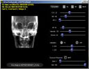
Click image to see a selection of screen shots produced by this module.
This module is different in operation. There are six datasets; Skull, C. Spine, Thorax, L. Spine, Pelvis, and Left Knee. It is suitable for installation in a variety of locations on the campus. It is intended for use with a projector working at 800x600 or greater screen resolution and 24 bit colour or greater. It is designed to provide real time changes to the radiographic image to support lectures and workshops with larger student groups.
It features: