shaderware recommend this title as an accompaniment to virtual radiographyTM
shaderware recommend this title as an accompaniment to virtual radiographyTM
Case study - School of Health and Social Care, University of Teesside, Middlesbrough, UK - http://www.tees.ac.uk/schools/SOH/
See the Promotional video for the School of Health and Social Care at The University of Teesside featuring virtual radiography
Watch a short video by Philip Cosson explaining how and why virtual radiography is used at The University of Teesside.
Number of students 40 undergraduate, 15 postgraduate
Program Leaders - Barbara Wilford and Susan Nixon
Date of adoption of Virtual RadiographyTM - February 2004
Current Version Installed Virtual RadiographyTM 3.0 (January 2006)
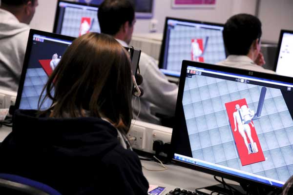
The diagnostic radiography course at Teesside moved into the university setting in 1992 from a hospital based education centre at James Cook University Hospital. At that time the live x-ray room in the education centre was still used and students were timetabled sessions in this environment. In 2000 the whole education centre was relocated, with the nursing and midwifery provision, to the university in a new building. The x-ray equipment was too old to transfer and the COSSH regulations concerning chemical waste meant the only way to re-provide a live x-ray room was to go digital. The decision was made not to do this. Instead, a new 20 seat computer lab with a database of 50,000 radiological images was provided. This lab is used extensively by all subject groups from social work to nursing, midwifery to physiotherapy. Software includes SPSS, Nvivio, DICOM viewers, Microsoft office, Bellwood (a virtual world for nurse education) and AiMs (radiological database).
Initially practical lab sessions in the local hospitals were planned but this proved difficult to timetable during normal working hours. In 2003, an x-ray room was installed in one of the clinical skills labs, but this is not live. It is used in role play during initial radiography specific modules. In 2004, Virtual RadiographyTM 2.0 was installed together with a computer upgrade. Evaluation fromstudents was very positive, and an upgrade to Virtual RadiographyTM 3.0 took place in 2006. All first year students now have 4 hours of lab time on virtual radiography prior to clinical placement. Second year students have a 3 hour workshop on skull and facial bones radiography during clinical placement to augment their experience (which is often completely absent). Some 3rd year radiographers have used virtual radiography as a research tool this year as part of their dissertation.
We mention we have virtual radiography in the marketing materials - applicants often wish to see the software during our open days; it causes interest Sally Abel, Admissions Officer
MSc students were the first to use it. The MSc is an accelerated course only taking 21 months from start to finish. The opportunity virtual radiography gives them to experiment, and find out for themselves what the effects of different changes are, really fits the problem based learning philosophy of the course Susan Nixon, Program Leader, MSc diagnostic radiography (preregistration)
The students are finding certain examinations rare in 9-5 practice, such as skull and facial bones. Students themselves requested an additional workshop, which we have put on for them in 2006 for the first time. The evaluation was good Sue Cutler, clinical coordinator.
Virtual radiography has proved simple to install and maintenance free. It coexists with all our preinstalled software and has survived a major upgrade of operating system with no difficulties. The lab is secured using Faronics DEEPFREEZE enterprise, and this causes no problems David Harris. IT Manager